Ventricular Tachycardia (VT)
Ventricular Tachycardia is a potentially lethal regular wide-complex tachycardia in which the ventricles have taken over rapid pacemaking of the heart.
As the name implies, in Ventricular Tachycardia the ventricles themselves become the origin of electrical signals and generate them at a tachycardic rate. Since these electrical signals arise from random ventricular cells, they are outside of the hearts specialized conduction system and propagate slowly across tissue. The hallmark of this slow propagation is a wide (longer) QRS complex.
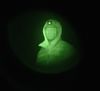
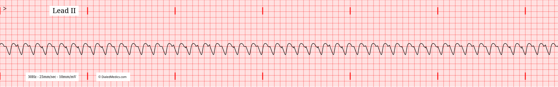
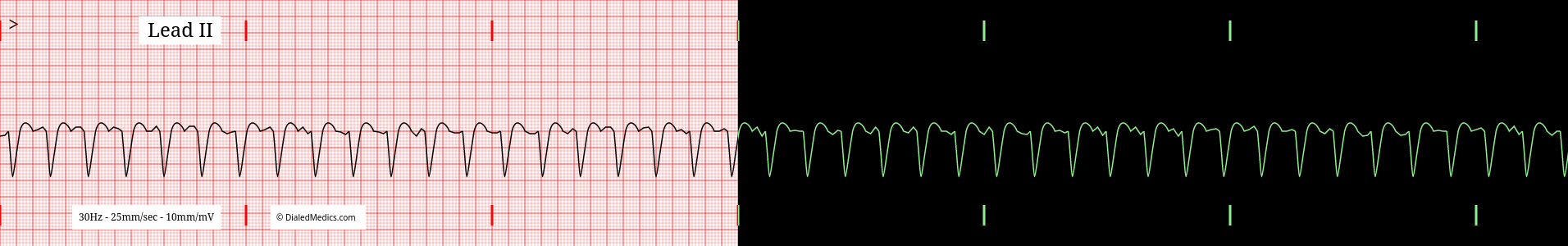
1:00 Ventricular Tachycardia monitor capture.
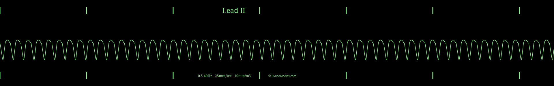
Once a tracing is identified as having a ventricular origin, wide QRS complexes, and a tachycardic rate, it can be called Ventricular Tachycardia. A Ventricular Tachycardia EKG is:
An EKG tracing showing a tachycardic rate with a ventricular origin (wide QRS Complex.)
Defining Characteristics of VTach in Lead II
- Rate: > 99 | Generally > 150.
- Rhythm: Regularly Regular | Equal R-R Intervals.
- P Waves: Not present | Non-sinus origin.
- P-R Intervals: N/A
- QRS Complexes: > 0.12sec (3mm) [3 small boxes] | Wide.
- Q-T Intervals†: Approx. 0.42sec (10.5mm) [10.5 small boxes].
- T Waves: Present after every QRS Complex.
† Q-T Intervals are variable with HR, age, and sex.
Rates of Ventricular Tachycardia
Being tachycardic, VTs rate must be at least 100bpm. As a generality, Ventricular Tachycardia tends to present at rates greater than 150bpm, but any rate over 100 qualifies.
Expected Rhythm in VTach
Ventricular Tachycardia is a regularly regular rhythm. It has a steady and even beat like a metronome.
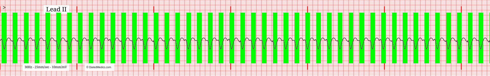
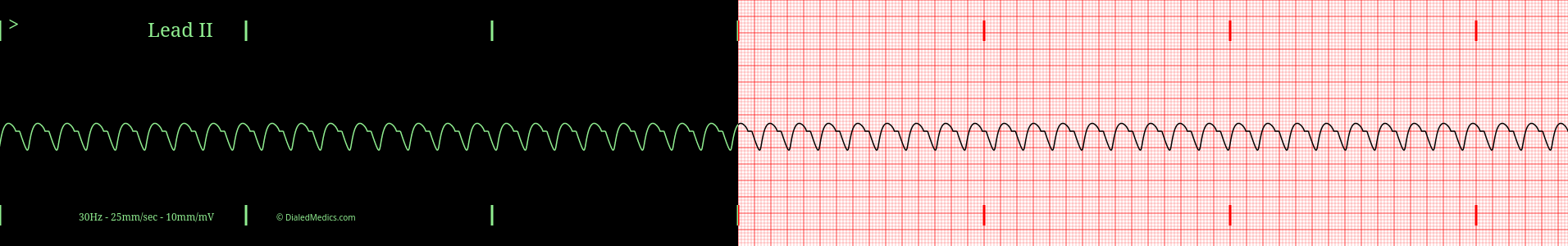
The QRS Complex in Ventricular Tachycardia
VT is a Wide Complex Tachycardia and as such is expected to have a wide (>12mm) [0.12sec] QRS complex. The specific morphology is dependent on exactly where in the ventricle the electrical signal is originating and will be different in every case. Below is an example tracing with wide QRS complexes highlighted in green.
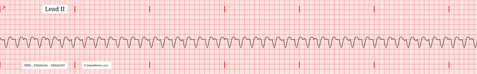
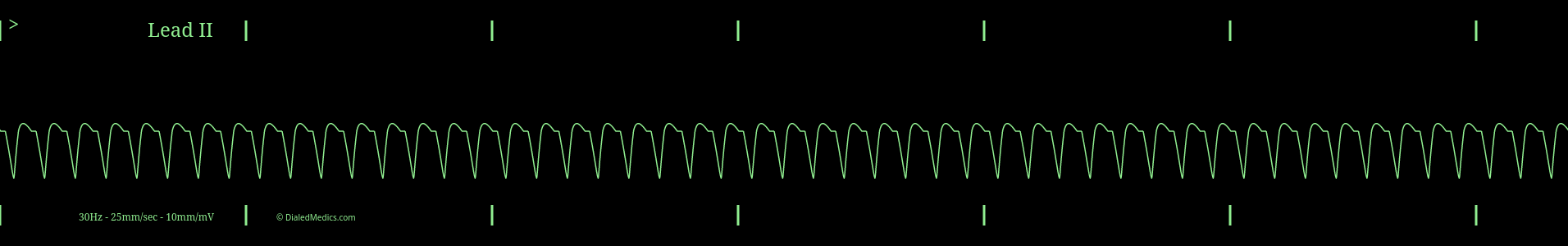
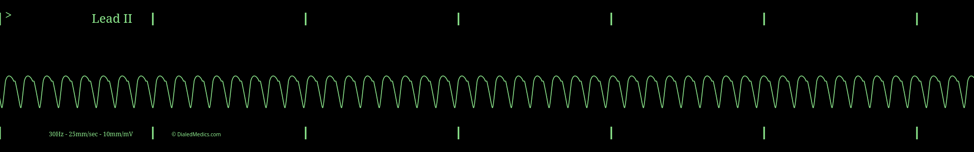
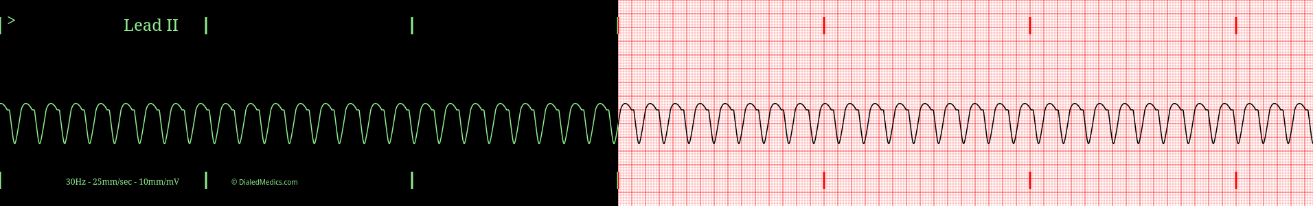
Below are several more examples of Ventricular Tachycardia presented as monitor captures as well as on EGK graphs.
After becoming confident in identifying EKGs as Ventricular Tachycardia head back to our EKG Rhythm Index to find information on another ECG. Otherwise practice interpreting novel EKGs with our EKG Generator:
Basic EKG App
Our Basic EKG Generator is free with an email signup and covers Normal Sinus Rhythm along with common arrhythmia.
Pro EKG App
Our Pro EKG Generator covers over 40 different rhythm categories, multiple display options, has Quiz and Simulation modes, and more! Try it out for just $5 for a month.