Supraventricular Tachycardia (SVT)
Supraventricular Tachycardia (SVT) is a category rather than a specific rhythm. SVTs are tachycardic rhythms which originate above the ventricles.
The Supra in Supraventricular Tachycardia is Latin for above and references the origin of the electrical signal above the ventricles. Tachycardia is any rhythm with a Heart Rate (HR) of 100bpm or more. It is important to note that SVT is not a specific cardiac rhythm, it is a category that includes all tachycardic rhythms that originate above the ventricles.
However, in clinical practice, the term Supraventricular Tachycardia is reserved for EKG tracings which are too fast to differentiate which specific origin is present. While a high HR and narrow QRS Complex (origin above the ventricles) are easy to recognize, SVT is defined by not being able to identify the specific origin. This is because, at high rates, waves become so close together that they can obscure one another.
The origin of an electrical signal is marked by the first feature of the repeating complex on an EKG. In order to recognize this, QRST complexes must be spaced far enough apart to expose details between them. As the HR increases, waves become overlapping, sometimes making it impossible to determine specifics.
The category of SVT includes rhythms such as Sinus Tachycardia, Ectopic Atrial Tachycardia, Multifocal Atrial Tachycardia, Atrial Flutter and Atrial Fibrillation with a Rapid Ventricular Response, Atrio-Ventricular and Atrio-Ventricular Nodal Reentry Tachycardias, Junctional Tachycardia, and more. The term SVT is used when the specific mechanism of the tachycardia cannot be determined.
You will hear clinicians talk about "looking for the underlying rhythm of an SVT" which is: attempting to further classify a Supraventricular Tachycardia into a specific cardiac rhythm. Some treatments for SVT (e.g. vagal maneuvers or adenosine) can cause a pause in the extremely fast rate allowing interpretation of the "underlying rhythm" and thereby guiding further treatment. See your clinical textbook for further information as interventions are out of the scope of this page.
Once a clinician has identified that an EKG does not have enough information visible to determine which specific tachycardia is present, the tracing can be called the more general Supraventricular Tachycardia. Supraventricular Tachycardia means:
A tachycardic ECG tracing with narrow QRS complexes (non-ventricular origin) on which there is not enough information to make a more specific interpretation.
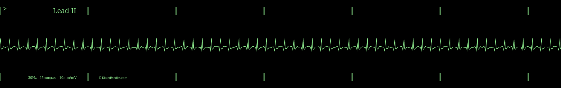
Defining Characteristics of SVT in Lead II
- RR Intervals less than 600ms (15mm) [3 large boxes] in duration.
- Less than 120ms (3mm) [3 small boxes] in duration.
* SVT can present with wide (> 120ms) QRS complexes. Such as when an SVT is present in conjunction with a bundle branch block or AV accessory pathway. These can result in Wide Complex Tachycardia, despite the supraventricular origin. However, there is no 100% sensitive and specific way to differentiate SVT with one of these types of aberrant conduction and Ventricular Tachycardia (VT). Therefore, Wide Complex Tachycardias should be assumed to be VT unless there is an extremely compelling clinical reason to consider SVT with aberrancy (such as a very young and otherwise healthy patient.) Differentiation cannot be attempted with a single lead and a 12-lead ECG will be required.
More important than meeting these criteria though is the absence of a clearly defined electrical origin.
The Rates of Supraventricular Tachycardia
As a tachycardia, SVT must have a rate of at least 100. In practice, only excessive rates of tachycardia obscure the origin of a cardiac rhythm, generally over 160. At rates below this the origin of a rhythm is usually visible between complexes and the specific Supraventricular Tachycardia (e.g. Sinus Tachycardia, Atrial Fibrillation with RVR, etc.) can be named.
While a specific rate isn't the determining factor of SVTs it is the high rate (frequent QRS complexes) which obscures other waveforms from interpretation.
QRS Complexes in SVT
The QRS complex in Supraventricular Tachycardia's are normal, as defined in the prototypical NSR:
- QRS Complexes: Maximum 0.12sec (3mm) [3 small boxes]
The narrow QRS complex of Supraventricular Tachycardia is confirmation of the supraventricular origin of the electrical signal. Electrical origins in the ventricles create wide QRS Complexes, so Narrow Complex Tachycardias are always of supraventricular origin.
The Unclear supraventricular Origin of SVTs
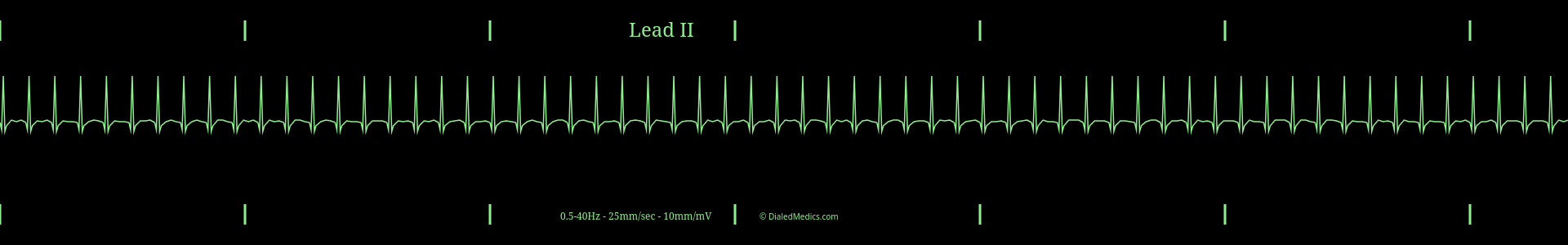
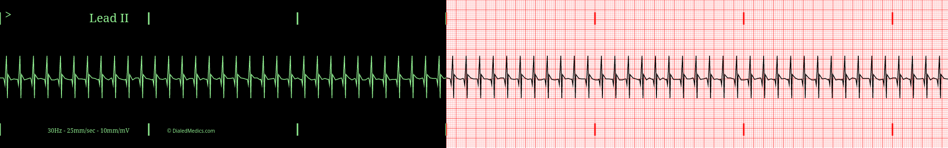
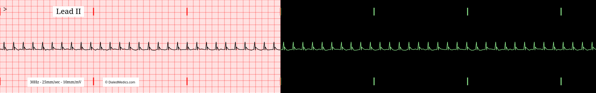
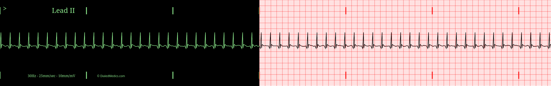
The key hallmark of Supraventricular Tachycardia is the indistinct origin. Narrow (< 0.12sec [3mm]) QRS Complexes indicate the origin above the ventricles, but the area where P Waves, fibrillation waves, or baseline would be is obscured by the end of the previous complex (marked in green above.)
The above tracing could be a Sinus Tachycardia, an Atrial Fibrillation with a Rapid Ventricular Response, or a Junctional Tachycardia, there is simply not enough information present to differentiate. This is the case when an EKG would be called only Supraventricular Tachycardia, until a specific underlying rhythm could be identified.
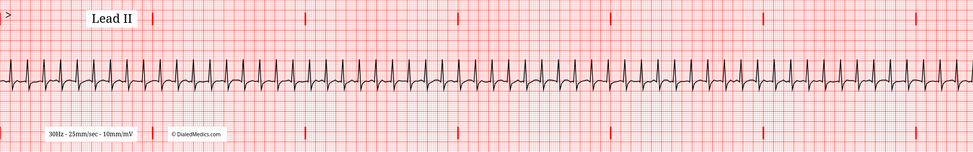
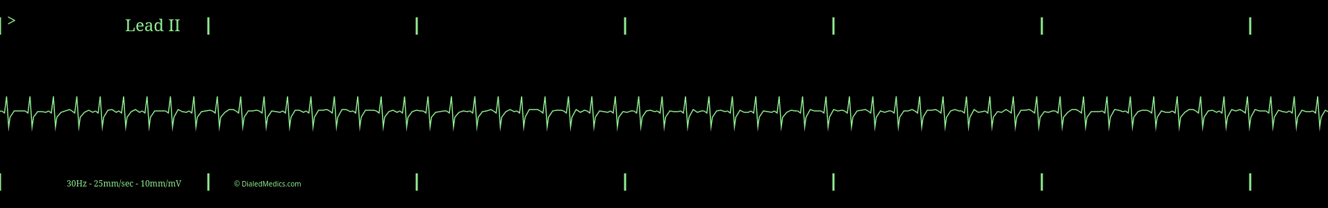
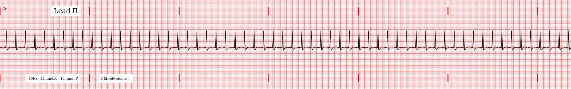
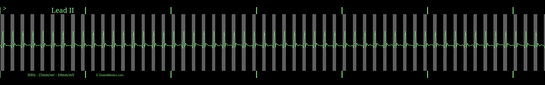
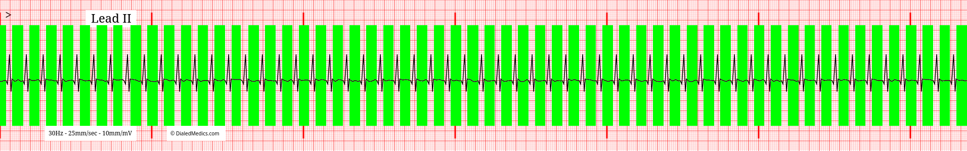
Below are several more examples of Supraventricular Tachycardia presented as monitor captures as well as on EGK graphs.
Once you're comfortable with the category of Supraventricular Tachycardia take a look at at Atrial Fibrillation or Sinus Tachycardia, two rhythms which may present as SVT when they have excessive rates. Or head back to our EKG Rhythm Index to find information on another ECG. Otherwise, practice interpreting novel EKGs with our EKG Generator:
Basic EKG App
Our Basic EKG Generator is free with an email signup and covers Normal Sinus Rhythm along with common arrhythmia.
Pro EKG App
Our Pro EKG Generator covers over 40 different rhythm categories, multiple display options, has Quiz and Simulation modes, and more! Try it out for just $5 for a month.