Atrio-Ventricular Paced Rhythm
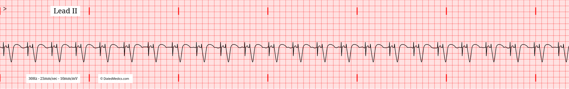
AV Pacemakers (a.k.a. Dual Chamber Pacing) artificially activates both the atria and ventricles in sequence. Sharp pacemaker spikes before both P waves and QRS complexes are indicative of AV pacing.
There are many types of pacemakers and many modes of pacemaker operation, this page is discussing specifically total sequential atrio-ventricular pacemakers. In these cases, the pacemaker has completely replaced natural pacemaking of the heart.
The hallmark of artificially paced EKG rhythms are pacer spikes. The electrical stimulus of the pacemaker results in a distinctive sharp wave which precedes atrial and then ventricular activation. In sequential atrio-ventricular pacing a single pacer spike will initiate each P Wave and QRS Complex, respectively.
Once a tracing is identified as having artificial pacer spikes initiating each wave it can be called a Paced Rhythm. A Total Atrio-Ventricular Pacemaker EKG is:
An EKG tracing with artificial pacer spikes which precede each P Wave and QRS complex in the tracing.
Pacer Spikes
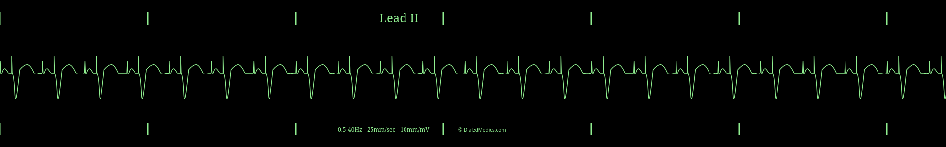
A hallmark of all paced rhythms are pacer spikes. Pacer spikes are short and sharp deflections which initiate the expected waves of the EKG tracing. Atrio-Ventricular sequential pacemakers will have two distinct pacer spikes per cycle; one for atrial activation (initial deflection) and one for ventricular deflection (second pacer spike) prior to the QRS Complex.
In the above example pacer spikes are marked in green. The initial pacer spike has a small positive deflection but is primarily a negative deflection and is marked with dark green arrows. This initial pacer spike is the atrial pacer spike, followed by the P Wave. The second pacer spike follows the P Wave and is marked with light green arrows. This ventricular pacer spike is followed by the QRS Complex.
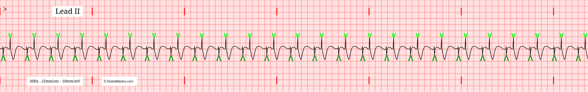
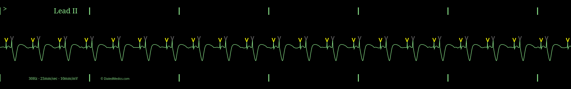
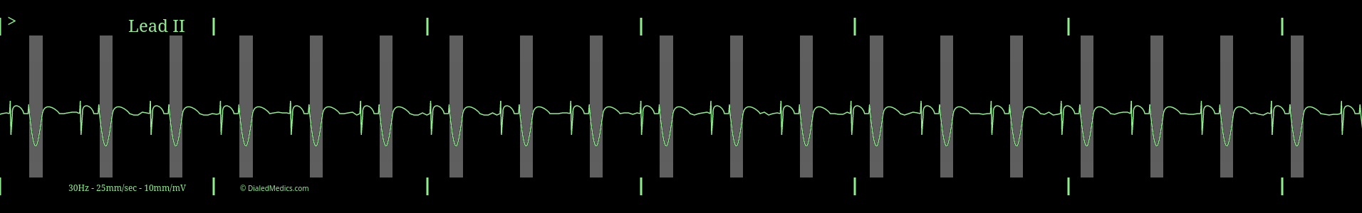
In the below example atrial pacer spikes have been marked in yellow and ventricular pacer spikes in grey.
Defining Characteristics* of Atrio-Ventricular Pacemakers in Lead II
- Rate: Artificially set ~70bpm.
- Rhythm: Regularly Regular | even steady beat like a metronome.
- P Waves: May be normal or Greater than 0.12sec (3mm) [3 small boxes] in duration.
- P-R Intervals: N/A
- QRS Complexes: Greater than 0.12sec (3mm) [3 small boxes] in duration.
- Q-T Intervals†: Approx. 0.42sec (10.5mm) [10.5 small boxes].
- T Waves: Present after wide QRS.
* Changes in paced rhythms may be an indication of pacemaker malfunction but don't ultimately change an EKGs categorization as a paced rhythm.
† Q-T Intervals are variable with HR, age, and sex.
Sequential AV Pacemaker Rate and Rhythm
The heart rate of a total sequential atrio-ventricular pacemaker is programatically set. In general the rate is around 70bpm, but this setting may have been adjusted to meet the cardiac output demands of any given PT.
Likewise, the rhythm is also programatically controlled and should be regularly regular.
Atrio-Ventricular Pacemaker Waveforms and Complexes
- P Waves: May be normal or Greater than 0.12sec (3mm) [3 small boxes] in duration.
- QRS Complexes: Greater than 0.12sec (3mm) [3 small boxes] in duration.
A pacemaker initiates electrical signals but can't replicate the electrical function of the heart fully. Natural pacemaking potentials travel from their origin through specialized conduction pathways rapidly, creating the normal duration of waves described in Normal Sinus Rhythm. Artificial pacemaker potentials don't; they propagate through all cardiac cells moving outward from the pacemaker electrode slowly relative to natural signals. As a result, the P Wave and QRS Complex of artificially paced rhythms are wider (longer) than expected.
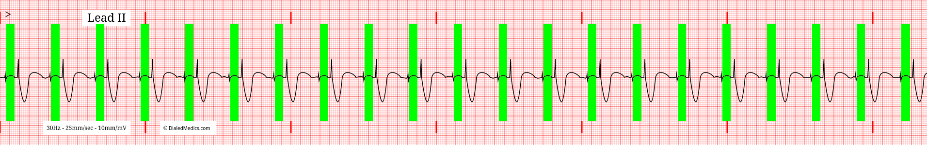
Above is an example of an Atrio-Ventricular Paced EKG with the wide P Waves highlighted in green. Note the atrial pacer spike initiating the wide P Wave. Below is a monitor capture of an AV Paced rhythm with its wide QRS complex highlighted in grey.
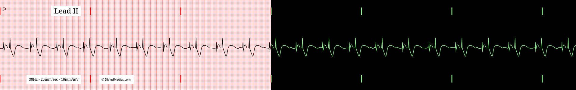
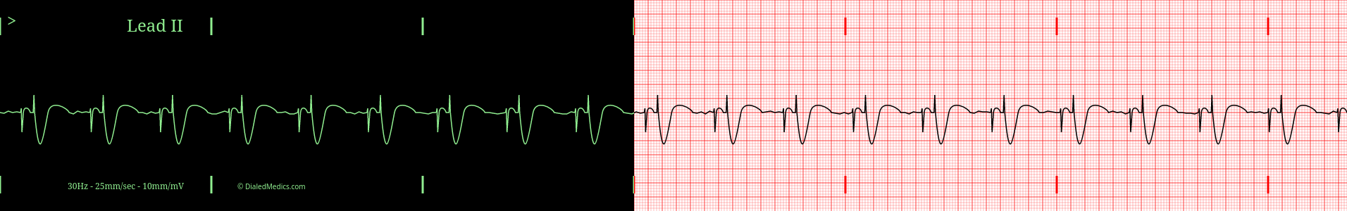
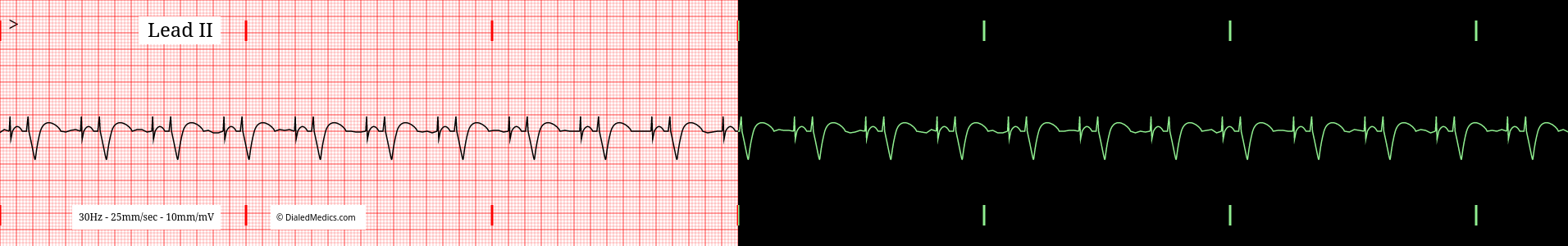
Below are several more examples of Total Sequential Atrio-Ventricular Pacemaker tracings presented as monitor captures as well as on EGK graphs.
After becoming confident in identifying EKGs as Total Sequential Atrio-Ventricular Pacemakers head back to our EKG Rhythm Index to find information on another ECG. Otherwise practice interpreting novel EKGs with our EKG Generator:
Basic EKG App
Our Basic EKG Generator is free with an email signup and covers Normal Sinus Rhythm along with common arrhythmia.
Pro EKG App
Our Pro EKG Generator covers over 40 different rhythm categories, multiple display options, has Quiz and Simulation modes, and more! Try it out for just $5 for a month